PET Scan
Vista offers patients the option of a positron emission tomography (PET) scan, which is an imaging test that uses a radioactive substance called a tracer to look for disease in the body.
A PET scan shows how organs and tissues are working. This is different than magnetic resonance imaging (MRI) and computed tomography (CT), which show the structure of and blood flow to and from organs.
Why the Test Is Performed
A PET scan can reveal the size, shape, position, and some function of organs. This test can be used to:
- Check brain function
- Diagnose cancer, heart problems, and brain disorders
- See how far cancer has spread
- Show areas in which there is poor blood flow to the heart
Several PET scans may be taken over time to check how well you are responding to treatment for cancer or another illness.
How the Test Is Performed
A PET scan uses a small amount of radioactive material, called a tracer, which is injected into a vein, most often on the inside of your elbow. The tracer travels through your blood and collects in organs and tissues. This helps the radiologist see certain areas of concern more clearly.
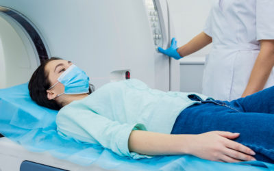
You will need to wait nearby as the tracer is absorbed by your body. This takes about an hour. Then, you will lie on a narrow table that slides into a large tunnel-shaped scanner. The PET up detects signals from the tracer while a computer changes the signals into 3-D pictures. The images are displayed on a monitor for your doctor to read.
How to Prepare For the Test
You may be asked not to eat anything for 4 – 6 hours before the scan, but you will be able to drink water.
We also can help you through this process if:
- You are afraid of close spaces (have claustrophobia). You may be given a medicine to help you feel sleepy and less anxious.
- You are pregnant or think you might be pregnant.
- You have any allergies to injected dye (contrast).
Always discuss the medicines you are taking, including those bought without a prescription. Sometimes, medications may interfere with the test results.
How the Test Will Feel
You may feel a sharp sting when the needle with the tracer is placed into your vein. A PET scan causes no pain. The table may be hard or cold, but you can request a blanket or pillow. An intercom in the room allows you to speak to someone at any time. There is no recovery time, unless you were given a medicine to relax.
Normal Results
A normal result means there were no problems seen in the size, shape, or position of an organ. There are no areas in which the tracer has abnormally collected.
Abnormal Results
Abnormal results depend on the part of the body being studied. Abnormal results may be due to: change in the size, shape, or position of an organ; cancer; infection; or problem with organ function.
Risks
The amount of radiation used in a PET scan is about the same amount as for most CT scans. Short-lived tracers are used so the radiation is gone from your body in about 2-10 hours.
Tell your doctor before having this test if you are pregnant or breast feeding. Infants and babies developing in the womb are more sensitive to radiation because their organs are still growing.
Rarely, people may have an allergic reaction to the tracer material. Some people have pain, redness, or swelling at the injection site.
Considerations
It is possible to have false results on a PET scan. Blood sugar or insulin levels may affect the test results in people with diabetes.
Most PET scans are now performed along with a CT scan. This combination scan is called a PET/CT.
For More Information
Call 847-360-6930
Related Services and Conditions
Digital Mammography
More than 200,000 new cases of breast cancer are discovered in the U.S. each year. At , we know the value of early detection and mammography is a tool we use to identify potential issues. An x-ray of the breast, mammography is used to look for...
Magnetic Resonance Imaging (MRI)
It’s clear—precision is important during diagnosis. Of all imaging technologies, MRI or Magnetic Resonance Imaging gives doctors the clearest, most precise image of the inside of the body. It’s sophisticated, and uses a strong magnetic field to show the structure and...
Diagnostic Imaging
At Vista Health System, we have board-certified technologists that capture quality images and ensure the comfort and safety of patients. These images are used to examine and diagnose certain medical conditions. Updated technology and diagnostic tools, including those...


